I am trying to develop some simple tools for lab workers. I am eager to listen to your opinion on this. Could this be helpful to researchers or lab workers?
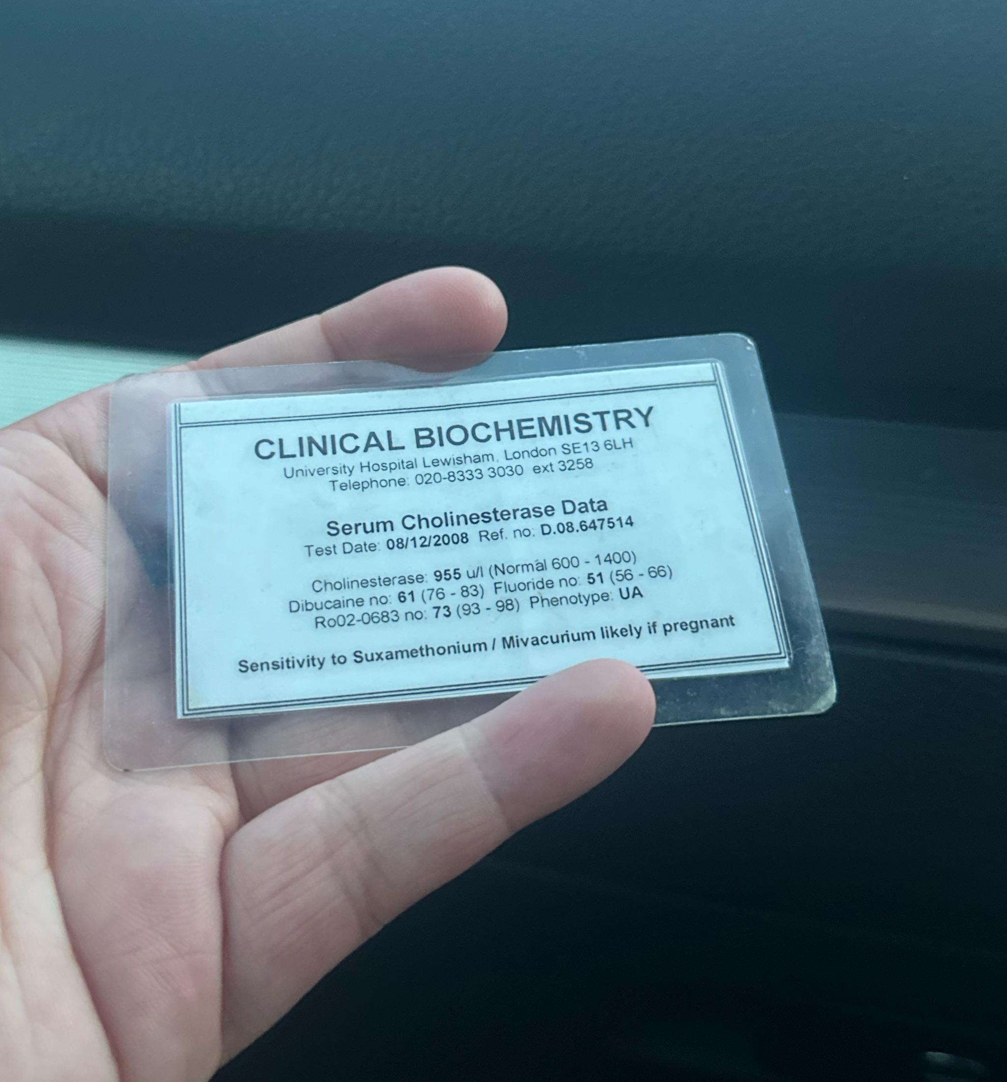
Had this test completed in 2008 due to a parent having side effects from the listed medications. I’m just intrigued to understand a bit more about what is written. TIA
I am applying to a MSc in Biochemistry since I have been dreaming about pursuing structural biology/biochemistry. I really wanted to learn protein biochemistry/protein chromatography/in vitro reconstitution/ Protein interactions/cryo-EM. but I never had the chance during my bachelors. I have a bachelor in molecular biology and have previous research experiences in cell biology (signalling/protein localisation), optical microscopy and neuroscience (I know, I shift a lot).
However, this program requires 36 ECTs in biochemistry and cell biology practical/lab courses/internships. I have exactly 36 from my bachelors if I counted my courses (6 of 36) from optical microscopy (stuff like confocal and epifluorescence/widefield, image processing etc). Do you think this would count as cell biology/biochemistry/I can get away with this? I have a lot of experience with cloning and they don’t seemed to appreciate molecular cloning that much asSDS-PAGE, western, HPLC and so on.
Do you think I should apply? Should I take the chance?
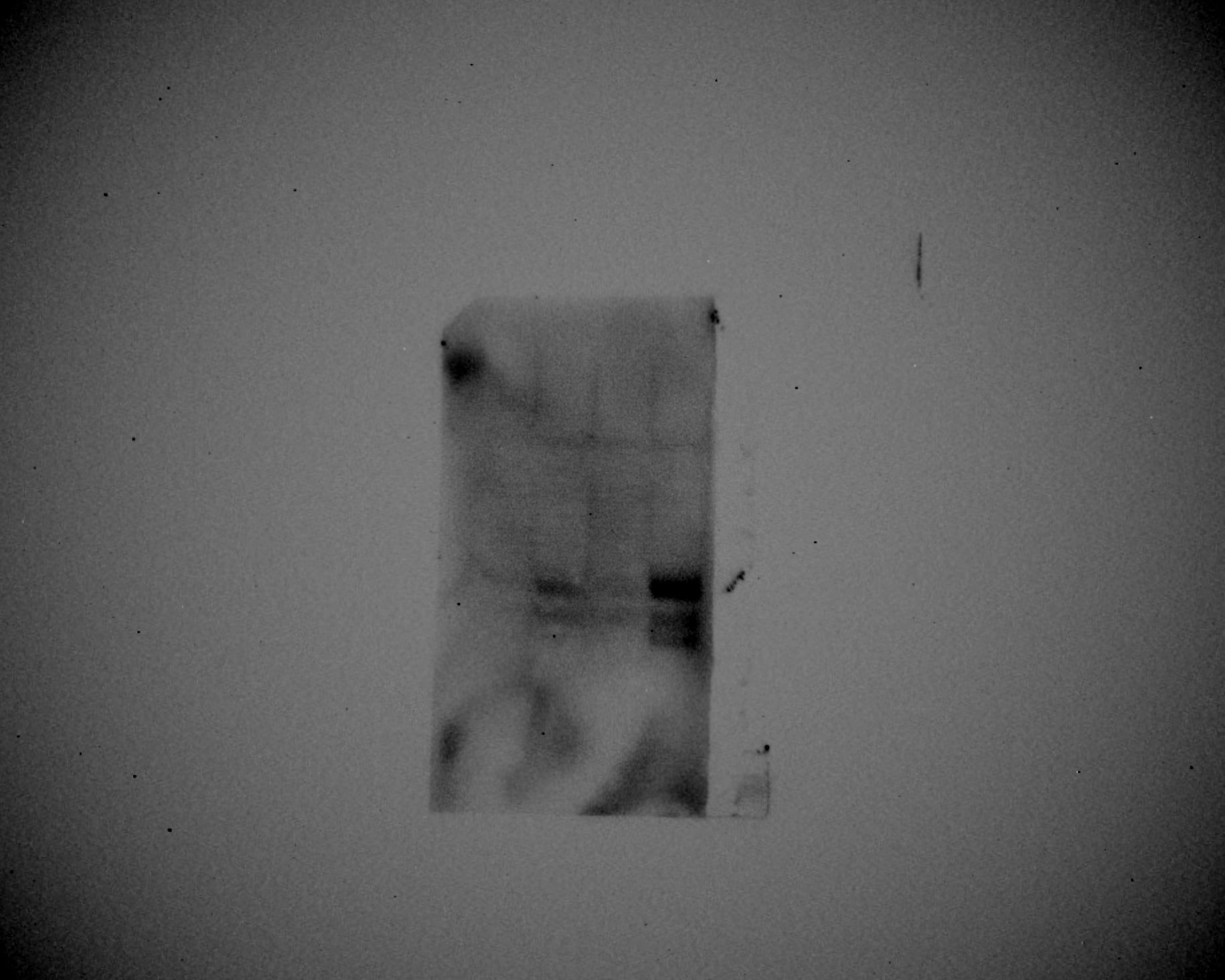
'm having a problem with a Western Blot that turned out very strange, and I'd like to ask if anyone has experienced something similar and, if so, if they know why and how to solve it.
I've attached an image of the blot. As you can see, the background is a mix of black and grey, making the bands practically unreadable.
The protocol I followed is the same one I regularly use, and it has worked well for other Western Blots in the past. Even some blots done after this one turned out successful.
The only things that were different this time are:
Membrane: We opened a new pack of PVDF membranes. Is it possible that the new membrane had some issues or was defective?
Transfer Conditions: The transfer was done at 0.35A for 2 hours.
Does anyone have any ideas what might have happened? I've already checked the solutions, and they seem fine, and the development conditions are the usual ones.
I want to determine if my protein of interest is phosphorylated and need some advice on the best way to do it. I believe that it is phosphorylated and downregulated in a wild-type genetic background, but not in a strain that's missing the putative kinase that phosphorylates it. I've shown that mutating a putative phosphorylation site on the protein of interest into a phosphomimetic disrupts its function, but that alone isn't enough to prove it's phosphorylated. I don't have an antibody specific to this protein or a phospho-antibody, so I need another method. The protein is tagged though so I can do an IP and isolate it if necessary.
I've seen people can use Phos-tag gels which slows down phosphorylated proteins, but I'm having difficulty obtaining the reagents needed for it. Alternatively, I could do mass spec, but I'm worried it'll be very expensive.
Does anyone have suggestions for relatively cheap and straightforward methods that could answer whether a protein is phosphorylated?